On-line: ISSN 2255–9582

| Faculty of Biology, University of Latvia | ||||||

|
Hard copy: ISSN 1691–8088
On-line: ISSN 2255–9582 Volume 18, Number 3
|
|||||

|
About the Journal | Retractions | Open Access | Author Guidlines | Current Issue | Archive |
|
Environmental and Experimental Biology |
Volume 18, Number 3 |
Balakrishnan S., Govindarajan R.
2020. Ultrastructural studies on the corticating filament of Chara zeylanica.
Environmental and Experimental Biology 18: 169–174.
DOI: 10.22364/eeb.18.17

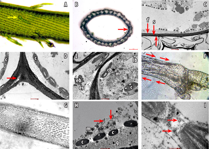
Fig. 1. Ultrastructure of Chara zeylanica cells. A, internodal cell with corticating filaments and spine cells (arrows). B, CS of internodal cell showing heavy calcification between the cell wall and corticating layer (arrows). C, cell wall of internode differentiated into outer fibrillar (f), middle spongy (s), inner fibrillar (i). Corticating cell with thick outer cell and thin inner cell wall closer to the cell wall of internodal cell. D, cell wall of two adjacent cortical cells showing charosome development. E, corticating cell with cell inclusions viz. chloroplast (c), lipid bodies (lb), endoplasmic reticulum (er), mitochondria (m), echinoid bodies (eb), and crystalline bodies (cb). F, Internodal cell under a light microscope showing cytoplasmic streaming (arrow). G, front view of an internodal cell showing uniform arrangement of chloroplast. H, chain of chloroplasts in the internodal cell with mitochondria (m), glycosomes (g) and endoplasmic reticulum (black arrow heads). I, microfilaments (arrows) running parallel between adjacent chloroplast.